Radiology

NEW GENERATION COMPUTED TOMOGRAPHY CAPABLE OF SPECTRAL IMAGING (256 SECTION DUAL ENERGY)

OPPORTUNITY FOR PATIENTS WITH PROSTHESIS

Patients with metal prostheses can be examined without artifacts,

NEW GENERATION CARDIAC CT APPLICATIONS

Perfusion examinations of cardiac muscles can be performed. In heart myocardial perfusion study, sections showing perfusion defects are color coded.

Contrast agents used during shooting can be virtually removed from the images without the need for extra shots. Contrast agents applied to patients are automatically detected by the system with numerical values in the tissues. Dangerous plaques hidden among lime plaques in the heart vessels can be visualized.

HIGH Spatial RESOLUTION

Situations where small structures such as coronary arteries need to be examined are one of the areas where systems must be pushed to their limits. In the CT system capable of performing spectral imaging used in our hospital, the spatial resolution of coronary vessel imaging is 18.2 lp/cm. The higher this value needed in cardiovascular stenosis examinations, the higher the image quality. The underlying technology that achieves this spatial resolution lies in the "Primary Speed" feature, which is 100 times more than traditional technology, and the "Afterglow" time, which is 4 times faster.

VIRTUAL ENDOSCOPY

Imaging of the large intestine for endoscopic examinations performed for screening purposes, where the interventional method is not mandatory, is possible with our CT system.

NEW GENERATION IT APPLICATIONS

It is possible to analyze the characterization of kidney stones according to their degree of hardness and determine the type of treatment. Contrast material retentions that cannot be distinguished visually can be analyzed numerically.

LOW DOSE, PATIENT SAFETY

X-ray safety, the system uses advanced patient safety technologies to use the lowest possible X-ray beam.

3D Dose Modulation allows body parts with different thickness and density to be shot with different X-ray doses in the same shot.
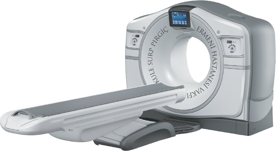
Tomografi Cihazı

Shooting protocols for all ages between 0-18 are provided to the user with maximum security. In the method called "Adaptive Statistical Iterative Reconstruction" (ASiR) technique, it is possible to obtain HD image quality CT images with much lower X-ray exposure than previous technologies.

HEART ATTACK RISK CALCULATION

The diagnosis of calcifications that narrow the heart vessels is made within seconds by CT examination and analysis (SmartScore) without even using contrast material. As a result of scoring analyses, which include factors such as patients' diabetic status, blood analysis, smoking and age, their risk of having a heart attack in the first ten years can be expressed as a percentage.

ADVANCED APPLICATIONS

Especially the damage and stenosis of the neck or brain vessels passing through the bone structures can be evaluated without bone structures.

In lung imaging, nodules are automatically tracked by the systems. Brain and liver perfusion imaging and analysis can be performed to monitor TM changes after radiotherapy in oncology cases and to diagnose stroke cases.

As a result, CT examinations, in which quantitative as well as qualitative values can be obtained with high accuracy and these results are taken into consideration during diagnosis, can be performed in the radiology department of our hospital.

TOMOSENTEZ DİJİTAL MAMOGRAFİ

We are monitoring your concerns in all their dimensions! Now mammography is much more comfortable and reliable. With the world's least irradiating Tomosynthesis Digital Mammography System, we can access extremely sharp images and finest details in three-dimensional mammography examination.

Early diagnosis is very important for a healthy future...

With this method, many images are taken from different angles and a 3D holistic image of the breast tissue is obtained on the computer with these images. Thus, cancers that can be overlooked due to overlapping tissues can now be revealed more clearly. This mammography device, which is the world's first patient-controlled remote compression control, puts compression control completely under the patient's control during imaging. Do not neglect your regular annual check-ups. Early diagnosis is very important for a healthy future…

Mamografi Cihazı
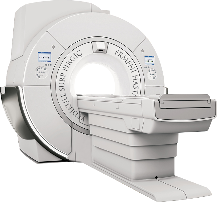
Tesla Sentetik MR

NEW GENERATION QUIET 3.0 TESLA SYNTHETIC MR

SILENT MRI

The "SILENT MRI", which leaves its mark on the patient imaging experience, is an important step taken for pediatric patients. This technology, which makes it possible to take images without anesthesia, especially for pediatric patients, brings superior image quality by eliminating image distortions caused by the patient's movement, thanks to the fast hardware design and software it is based on.

WIDE PATIENT TUNNEL

70 cm wide tunnel opening, wider patient table, more spacious lighting and internal ventilation systems, lighter and more flexible surface coils and advanced software that can complete multiple imaging operations in a single shot in a short time...

NEEDLE-FREE APPLICATIONS

Within the scope of the patient-friendly "Needle-Free Applications" concept, the ability to perform MR angiography without injecting contrast material (drug) into the patient constitutes an important alternative for imaging especially pregnant, pediatric patients and patients with kidney failure.

Thanks to the new generation software within the scope of the patient-friendly "Needle-Free Applications" concept, fewer biopsy procedures are needed to make a diagnosis.

DESIGN THAT PROMOTES PATIENT COMFORT

Thanks to the superficial coils and patient table designed with patient comfort in mind, imaging of obese patients is now much more comfortable... Thanks to this design, all MRI scans can be performed either head first or foot first into the patient tunnel.

BREATHLESS-FREE SHOTS

Thanks to new generation software, it is now possible for patients who have difficulty holding their breath during MRI to have MRI scans without holding their breath.

NEW GENERATION SOFTWARE

In cases of stroke and microbleeding, early diagnosis saves lives with our MRI device, which includes advanced neurology software.

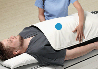
AİR COİL (MR Battaniyesi)

MR Battaniyesi

In the old system Magnetic Resonance imaging technology (for example) in Lung and Abdominal MRI examinations, the coils were adjusted according to the body region.

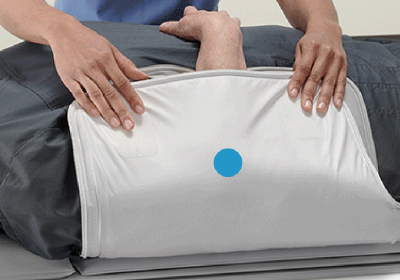
MR Battaniyesi

Each region was scanned separately with these procedures, which sometimes required moving the patient. With this state-of-the-art blanket, the shots are taken twice and completed in a single procedure, without moving the patient.

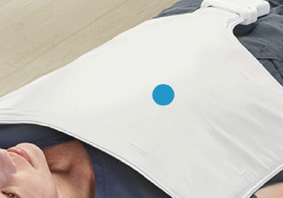
MR Battaniyesi

Because instead of spiral scanners produced with specific and inflexible molds, there are now devices that are soft and hug the body, like a blanket, and can scan larger surfaces at once.

MR Battaniyesi

Moreover, it is possible to achieve faster, more detailed and more reliable results with these devices. Coils are one of the basic components that provide image formation in MRI.

Premium Level Color Doppler Ultrasound

Combined with the dedicated biopsy probe and the newly expanded E Series probe family, the S-Agile architecture excels across a broad suite of clinical applications including abdominal, musculoskeletal, small organ, breast, vascular, obstetrics and gynecology, cardiology and more. It helps to achieve image quality.

Ultrason Cihazı

Consult a Doctor

This field is required.